Effect of calcination treatment on the in vitro bioactivity of ceramic powders
 View 45/
 12
0
12
0
DOI:
https://doi.org/10.69717/jaest.v6.i3.168Keywords:
Calcium phosphate, Calcination, Bioactivity, Physiological solution, Bioceramic powder, Tissue engineering, Calcium phosphate, Calcination, Bioactivity, Physiological solution, Bioceramic powder, Tissue engineeringAbstract
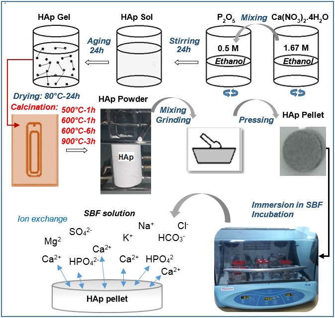
The aim of this study was to evaluate the effect of varying calcination temperature and duration (500°C -1h, 600°C-1h, 600°C-6h, and 900°C-3h) on the bioactivity property of crystalline hydroxyapatite pellets immersed in a physiological solution (simulated body fluid) for 7 days. The microstructural properties and composition of the powders−such as the degree of crystallinity, crystallite size, additional thermal phases of -tricalcium phosphate (β-TCP) and CaO, and functional groups (hydroxyl and carbonate)−vary depending on the calcination parameter and immersion process. X-ray diffraction (XRD) and infrared spectroscopy (IR) analyses showed significant microstructural changes for the HAP calcined at 900°C for 3 hours. Moreover, atomic absorption spectroscopy (AAS) analysis and pH measurements indicated variations in calcium ion concentration (42.2 ± 0.70 mg/L after 3 days, and 34.2 ± 0.48 mg/L after 7 days of immersion time) and pH of the medium-which decreased from 7.440 ± 0.02 to 7.305 ± 0.03-for the HAP pellet calcined at 900°C. These results were confirmed by X-ray fluorescence (XRF) and electron microscopy, which indicated an increase in the Ca content from 55.06 ± 0.156% to 55.85±0.148%, and in the P content from 24.64±0.155% to 27.59±0.158%, as well as the deposition of dispersed particles and large agglomerates, suggesting the formation of a non-uniform layer of calcium phosphate (CaP) on the surface of the powder pellet calcined at 900°C.
Highlights
- Variations in the calcination parameters of hydroxyapatite affect its biological response.
- CaP deposit formation after 7 days of immersion on the surface of HAp calcined at 900°C.
- The thermal decomposition of HAp can promote the formation of CaP deposits.
Downloads
References
A. Magnaudeix, Calcium phosphate bioceramics: From cell behavior to chemical-physical properties, Frontiers in Biomaterials Science 1 (2022) 942104. https://doi.org/10.3389/fbiom.2022.942104
S. Yala, M. Boustta, O. Gallet, M. Hindié, F. Carreiras, H. Benachour, D. Sidane, H. Khireddine, New synthesis method of HA/P(D,L)LA composites: Study of fibronectin adsorption and their effects in osteoblastic behavior for bone tissue engineering, Journal of Materials Science: Materials in Medicine 27 (2016) 140. https://doi.org/10.1007/s10856-016-5756-8
D. Sidane, H. Khireddine, S. Yala, The hardness of the hydroxyapatite-titania bilayer coatings by microindentation and nanoindentation testing, Journal of Physics: Conference Series 939 (2017) 012027. https://doi.org/10.1088/1742-6596/939/1/012027
D.F. Fitriyana, F.W. Nugraha, M.B. Laroybafih, R. Ismail, A.P. Bayuseno, R.C. Muhamadin, M.B. Ramadan, A.R.A. Qudus, J.P. Siregar, The effect of hydroxyapatite concentration on the mechanical properties and degradation rate of biocomposite for biomedical applications, IOP Conference Series: Earth and Environmental Science 969 (2022) 012045. https://doi.org/10.1088/1755-1315/969/1/012045
X. Yuan, T. Wu, T. Lu, J. Ye, Comparison of physicochemical and biological properties of calcium phosphate cements incorporating different strontium-containing compounds, Ceramics International 51 (2025) 60094–60105. https://doi.org/10.1016/j.ceramint.2025.10.208
M.G. Raucci, D. Giugliano, L. Ambrosio, Fundamental properties of bioceramics and biocomposites, in: I. Antoniac (Ed.), Handbook of Bioceramics and Biocomposites, Springer, Cham, 2015.
S. Ferraris, S. Yamaguchi, N. Barbani, M. Cazzola, C. Cristallini, M. Miola, E. Vernè, S. Spriano, Bioactive materials: In vitro investigation of different mechanisms of hydroxyapatite precipitation, Acta Biomaterialia 102 (2020) 468–480. https://doi.org/10.1016/j.actbio.2019.11.024
J. Wang, L. Zhang, K. Wang, Bioactive ceramic-based materials: Beneficial properties and potential applications in dental repair and regeneration, Regenerative Medicine 19 (2024) 257–278. https://doi.org/10.1080/17460751.2024.2343555
S. De Micco, D. Bellucci, V. Cannillo, The assessment of bioactivity and biological responsiveness in bioactive glasses and ceramics: A review of available techniques, Materials 18 (2025) 4393. https://doi.org/10.3390/ma18184393
P.A. Forero-Sossa, J.D. Salazar-Martínez, A.L. Giraldo-Betancur, B. Segura-Giraldo, E. Restrepo-Parra, Temperature effect in physicochemical and bioactive behavior of biogenic hydroxyapatite obtained from porcine bones, Scientific Reports 11 (2021) 11069. https://doi.org/10.1038/s41598-021-89776-2
T. Kokubo, H. Takadama, How useful is SBF in predicting in vivo bone bioactivity? Biomaterials 27 (2006) 2907–2915. https://doi.org/10.1016/j.biomaterials.2006.01.017
D. Sidane, H. Khireddine, Microstructural modification of calcium phosphate-based bioceramic materials by adding gelatin, Asia-Pacific Journal of Chemical Engineering 20 (2025) e70075. https://doi.org/10.1002/apj.70075
T. Kokubo, Bioceramics and Their Clinical Applications, Woodhead Publishing Limited, UK, 2008.
M. Furko, Bioglasses versus bioactive calcium phosphate derivatives as advanced ceramics in tissue engineering: Comparative and comprehensive study, current trends, and innovative solutions, Journal of Functional Biomaterials 16 (2025) 161. https://doi.org/10.3390/jfb16050161
H.M. Kim, T. Himeno, T. Kokubo, T. Nakamura, Process and kinetics of bonelike apatite formation on sintered hydroxyapatite in a simulated body fluid, Biomaterials 26 (2005) 4366–4373. https://doi.org/10.1016/j.biomaterials.2004.11.022
A. Singh, Hydroxyapatite, a biomaterial: Its chemical synthesis, characterization and study of biocompatibility prepared from shell of garden snail, Helix aspersa, Bulletin of Materials Science 35 (2012) 1031–1038. https://doi.org/10.1007/s12034-012-0384-5
A. Alshemary, M. Akram, Y. Goh, U. Tariq, F. Butt, A. Abdolahi, R. Hussain, Synthesis, characterization, in vitro bioactivity and antimicrobial activity of magnesium and nickel doped silicate hydroxyapatite, Ceramics International 41 (2015) 11886–11898. https://doi.org/10.1016/j.ceramint.2015.06.003
Z.H. Cheng, A. Yasukawa, K. Kandori, T. Ishikawa, FTIR study on incorporation of CO2 into calcium hydroxyapatite, Journal of the Chemical Society, Faraday Transactions 94 (1998) 1501–1505. https://doi.org/10.1039/A708581H
D.G. Chukhchin, A.V. Malkov, I.V. Tyshkunova, L.V. Mayer, E.V. Novozhilov, Diffractometric method for determining the degree of crystallinity of materials, Crystallography Reports 61 (2016) 371–375. https://doi.org/10.1134/S1063774516030081
S. Mondal, S. Park, J. Choi, T.T.H. Vu, V.H.M. Doan, T.T. Vo, B. Lee, J. Oh, Hydroxyapatite: A journey from biomaterials to advanced functional materials, Advances in Colloid and Interface Science 321 (2023) 103013. https://doi.org/10.1016/j.cis.2023.103013
H. Wang, J.K. Lee, A. Moursi, J.J. Lannutti, Ca/P ratio on the degradation of hydroxyapatite in vitro, Journal of Biomedical Materials Research Part A 67 (2003) 599–608. https://doi.org/10.1002/jbm.a.10538
A.D. French, M. Santiago Cintrón, Cellulose polymorphy, crystallite size, and the Segal crystallinity index, Cellulose 20 (2013) 583–588. https://doi.org/10.1007/s10570-012-9833-y
D. Predoi, C.S. Ciobanu, S.L. Iconaru, K. Rokosz, S. Raaen, S.A. Predoi, S. Ţălu, M. Motelica-Heino, Comprehensive physicochemical and biological analysis of hydroxyapatite/dextran powders before and after immersion in Kokubo solution, Langmuir 41 (2025) 17011–17034. https://doi.org/10.1021/acs.langmuir.5c01585
I. Galan, F.P. Glasser, C. Andrade, Calcium carbonate decomposition, Journal of Thermal Analysis and Calorimetry 111 (2013) 1197–1202. https://doi.org/10.1007/s10973-012-2290-x
A. Costescu, I. Pasuk, F. Ungureanu, A. Dinischiotu, M. Costache, F. Huneau, S. Galaup, P. Le Coustumer, D. Predoi, Physico-chemical properties of nano-sized hexagonal hydroxyapatite powder synthesized by sol-gel, Digest Journal of Nanomaterials and Biostructures 5 (2010) 989–1000.
S.A. Siddiqi, U. Azhar, Carbonate substituted hydroxyapatite, in: A.S. Khan, A.A. Chaudhry (Eds.), Handbook of Ionic Substituted Hydroxyapatites, Woodhead Publishing Series in Biomaterials, 2020, pp. 149–173. https://doi.org/10.1016/B978-0-08-102834-6.00006-9
C. Combes, C. Rey, Amorphous calcium phosphates: Synthesis, properties and uses in biomaterials, Acta Biomaterialia 6 (2010) 3362–3378. https://doi.org/10.1016/j.actbio.2010.02.017
H.M. Kim, T. Himeno, M. Kawashita, T. Kokubo, T. Nakamura, The mechanism of biomineralization of bone-like apatite on synthetic hydroxyapatite: An in vitro assessment, Journal of the Royal Society Interface 1 (2004) 17–22. https://doi.org/10.1098/rsif.2004.0003
A. Kurzyk, A. Szwed-Georgiou, J. Pagacz, et al., Calcination and ion substitution improve physicochemical and biological properties of nanohydroxyapatite for bone tissue engineering applications, Scientific Reports 13 (2023) 15384. https://doi.org/10.1038/s41598-023-42271-2
D. Shi, G. Jiang, J. Bauer, The effect of structural characteristics on the in vitro bioactivity of hydroxyapatite, Journal of Biomedical Materials Research 63 (2002) 71–78. https://doi.org/10.1002/jbm.10087
Published
Issue
Section
License
Copyright (c) 2026 Djahida Sidane, Nadir Slimani, Yasmina Bourebaba, Nawal Deflaoui, Lynda Sadou, Hafit Khireddine (Author)

This work is licensed under a Creative Commons Attribution 4.0 International License.
The Journal of Applied Engineering Science and Technology (JAEST) provides immediate open access to its content on the principle that making research freely available to the public supports a greater global exchange of knowledge.
All articles published in JAEST are licensed under the Creative Commons Attribution 4.0 International License (CC BY 4.0). This license permits users to share, copy, redistribute, adapt, and reproduce the published material in any medium or format, including for commercial purposes, provided that appropriate credit is given to the original author(s) and source, a link to the license is provided, and any changes made are indicated.
Authors retain the copyright of their published work and grant JAEST the right of first publication. Copyright is not transferred to the journal or publisher.
License: Creative Commons Attribution 4.0 International (CC BY 4.0)
License URL: Creative Commons CC BY 4.0